Summary
The article originally titled “Human organoid model of PCH2a recapitulates brain region-specific pathology” shows for the first time how the pathology in certain brain regions in PCH2A can be simulated in a 3-dimensional neuronal tissue model (organoid). With the help of this model of a brain affected by PCH2A, it is now possible to continue with intensive research and better understand the mechanisms of the disease.
Baseline
The genetic cause of PCH2A is a variation in the TSEN54 gene. However, the mechanisms that lead to cerebellar hypoplasia and progressive microcephaly are still unknown.
The main reason for this is that there is currently no adequate model for the disease.
What Is an Organoid?
However, progress has recently been made in the creation of organ models, so-called organoids. They can be thought of as a miniature version of an organ that has similar properties to a real human organ.
Creation of PCH2 Organoids
The research team under Kagermeier et al. took advantage of this to produce a human brain model for further research into PCH2A.
For this, they collected skin cells from three male PCH2 patients and used them to produce induced pluripotent stem cells (iPSCs). These cells are derived from body cells and then modified in such a way that they can multiply and mature into any type of human cell.
These iPSCs were then used to produce the organ models, specifically an organoid of the cerebellum and a so-called neocortical model (part of the cerebrum).
PCH2 Organoids Compared to the Control Group
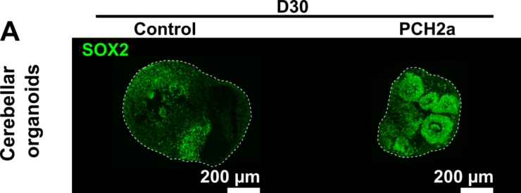
A comparison with organ models created from healthy cells, i.e. the control group, revealed, among other things, that the cerebellar organ models with PCH2 were significantly smaller.
The same was true for the neocortical models, but to a lesser extent than for the cerebellar models.This is reflected symptomatically in the increasingly reduced head circumference of the patients.
No Evidence of Cell Death in PCH2 Organoids
However, no evidence of apoptosis (cell death) was found in the organ models. This had been suspected in earlier studies based on animal models and pathological findings, but could not be confirmed. However, the organoids represent a much earlier stage of brain development than pathological studies have previously been able to investigate.
Another possible explanation could be that the precursor cells do not divide properly. The difference in size between PCH2a organoids and control organoids would then arise because not enough cells (including nerve cells) are formed. In brain organoids, these precursor cells are organized in round zones, so-called rosettes. This is where the majority of cell divisions take place. Detailed examinations of these rosettes in PCH2a cerebellar organoids revealed a difference to organoids of healthy subjects. This difference was not observed in the cerebral organoids. It can therefore be assumed that the reduced size of the cerebellum in PCH2a is at least partly due to deviations during brain development.
Conclusion
With the help of this model of a brain affected by PCH2A, it is now possible to continue with intensive research and better understand the mechanisms of the disease.
Read the Complete Study
Find the complete study here:


